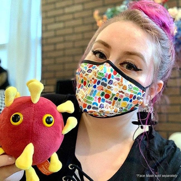


Antagligen det mest dödliga som finns där ute! Ett gossedjur med en förskräcklig historia. Men dock i mjuk och gosig plysch.
För varje såld Ebola stor mikrob skänker vi 25kr till hjälpinsatser mot Ebola i Västafrika.
Since its discovery in 1976, Ebola has become the T. Rex of microbes. Share the love!
GiantMicrobes är mjukdjur som ser ut som små, små mikrober - bara det att de är förstorade sådär en miljon gånger. De ger bokstavligen ett ansikte åt förkylningen, fotsvampen, hostan, den dåliga andedräkten eller vägglusen. Varje GIGANTmicrobe är mellan ca 38-50 cm. Med följer ett foto på hur den riktiga mikroben ser ut samt en kort information på engelska.
Ursprungligen skapade i USA att användas i undervisningssyfte, har GIANTmicrobes nu blivit storsäljare i museishoppar, apotek, bokhandlar och designbutiker världen över.
GIANTmicrobes är ett roligt verktyg i undervisningen om hälsa och sjukdomar, men även en uppskattad gåva som passar alla åldrar. Definitivt roligare än ett krya-på-dig kort till en sjuk vän.
--------------
Ebolavirus
| Ebola virus (EBOV) | |
|
Ebolavirus i mikroskop
|
|
| Mononegavirales | |
| Filoviridae | |
| Ebolavirus | |
| Bundibugyo ebolavirus, Zaire ebolavirus, Sudan ebolavirus, Reston ebolavirus, Côte d'Ivoire ebolavirus | |
Ebolavirus är det virus som orsakar ebolafeber, hemorragisk feber som är en blödarfeber med hög dödlighet. Viruset tillhör familjen Filoviridae och är ett stavformat filamentöst negativt enkelsträngat RNA-virus (-ssRNA.) Ebolavirus är ett zoonotiskt virus som kan smitta människor och djur via blod och kroppsvätskor. Det finns fem kända arter av ebolavirus; Zaire-, Sudan-, Côte d'Ivoire-, Reston- och Bundibugyo ebolavirus.[1] Ebolavirus är en av de mest dödliga patogenerna för människor och icke-humana primater och måste handhas på de laboratorier som har den högsta skyddsklassen, skyddsklass 4. Viruset angriper främst endotelceller och sprids via blodet ut i kroppen och orsakar nekros av kroppens celler, som leder till ett häftigt sjukdomsförlopp karakteriserat av blödningar ur kroppsöppningarna. Dödligheten är mellan 50-90%.[2] Till familjen Filovirus, Filoviridae hör förutom släktet ebolavirus, också släktet marburgvirus som även det orsakar hemorragisk feber.
Innehåll
[göm]- 1 Historia
- 2 Virusets uppbyggnad
- 3 Livscykel
- 4 Symptom
- 5 Behandling
- 6 Referenser
Historia[redigera | redigera wikitext]
De första dokumenterade fallen av ebolafeber hos människor noterades i Zaire och fick namnet efter floden Ebola. Utbrottet skedde 1976 då 318 människor insjuknade. Av dessa dog ca 90% och detta är den högsta mortalitetsration som noterats. Detta utbrott var av arten Zaire ebolavirus. De kända utbrotten har skett i centrala Afrika. Undantagen från detta har varit olyckor i laboratoriemiljö. Ebolavirus av arten Reston har även påträffats hos apor med ursprung i Filippinerna, men har då endast drabbat apor. Ebola hemmorragisk feber sprids troligtvis av fladdermöss som via spillning och kroppsvätskor kan sprida viruset vidare till människor och apor, såsom Gorillor och Schimpanser via bland annat djurkadaver. Virusets förmåga att infektera människor innebär sålunda att viruset har en zoonotisk kapacitet.[3] Begravningsceremonier där de sörjande kommit i direkt kontakt med den avlidne samt överföring av virus när anhöriga vårdar en ebolainfekterad människa tros vara betydande anledningar till spridning av sjukdomen
Virusets uppbyggnad[redigera | redigera wikitext]
Ebolaviruset är en icke-cellulär organism som orsakar blödarfeber. Genomet är nukleinsyra i form av en negativ enkelsträngad, RNA molekyl (-ssRNA). När viruset befinner sig utanför en cell kallas det för virion och består då av en spiralformad proteinkapsel som omsluter genomet. Utanför cellen har inte viruset någon egen metabolism. Men när viruset tar sig in i en värdcell tar den kontroll över cellens metabolism och tillverkar mer virus.[4] Ebolavirusets membran består av kapsomerer som är bundna till varandra och på så sätt formar ett spiralformat rör som kan vara grenat, cirkulärt, u-format eller format som en 6:a.[5] Ebola vironets längd är ~805 nm.[6]
Livscykel[redigera | redigera wikitext]
Viruset kan inte reproducera sig självt utan måste ha hjälp av sin värdcell för att kunna föröka sig. Virusets övertagande av en cell innebär att värdcellen till slut lyserar. Filovirusets replikationscykel börjar med att viruset fäster till värdcellen med hjälp av en receptor och sedan endocyterar viruset genom membranet genom att aktinfilament binder in viruset till värdcellen genom macropinocytos [7]. För att syntetisera protein måste –ssRNA viruset bära med sig enzymet, RNA beroende RNA transkriptas. Detta för att –RNA inte känns igen av ribosomerna och därför måste översättas till +RNA (mRNA). Viruset syntetiserar proteiner och nukleinsyra med hjälp av värdcellens enzymer och ribosomer. De nya vironerna ansamlas i värdcellen varpå de nya vironerna exocyterar från värdcellen och livscykeln repeteras.[4] Ebolaviruset attackerar främst endotelceller. Vid infektion fäster virusets glykoproteiner till endotelceller som transporterar ut viruset i kroppen via blodet och orsakar infektion och nekros av celler i lymfkörtlar, lever, mjälte och lungor.[8][9]
Symptom[redigera | redigera wikitext]
Inkubationstiden för ebolafeber är mellan 2-21 dagar [10]. Symptomen är akut feber, huvudvärk, muskelvärk, torrhet i svalget, svaghet följt av yrsel, blodiga kräkningar och diarréer och blödningar ur kroppsöppningarna.[1][11] Diagnostiseringen sker med hjälp av ELISA (Enzymkopplad immunadsorberande analys), PCR (Polymeraskedjereaktion)eller genom virusisolation från blodprov. Under senare delen av sjukdomsförloppet eller under tillfrisknandet kan test av antikroppar göras. Diagnostiseringen sker ofta efter full utveckling av ebolafeber då de tidiga symptomen liknar många andra sjukdomar.[1][10]
Behandling[redigera | redigera wikitext]
Det saknas vaccin och antiviralt medel mot ebolafeber. Symptomen behandlas genom att ge patienten vätskeersättning och elektrolyter, febernedsättande läkemedel, vidbehålla patientens syrenivå och blodtryck. Ett antal vacciner har testats under åren men med begränsad framgång. Ribavirin har haft visad framgång bland infekterade möss, men ännu har det inte gjorts några behandlingar av människor med detta läkemedel. Vid upptäckt av Ebola-infektioner finns strikta riktlinjer och manualer utfärdade av WHO att följa. I riktlinjerna finns åtgärder som, kontaktspårning för att se hur många andra som den smittade varit i kontakt med, isolering av patienten, hygienregler som kräver komplett skyddsutrustning och att allt som varit i kontakt med patienten så som instrument och liknande desinficeras och förbrukningsmaterial destrueras. Ebola hemmoragisk feber är nivå 4-klassad, vilket är den högsta säkerhetsnivån i ett laboratorium, samt tillhör kategori A som potentiellt medel för användning inom bioterrorism.[1]
Referenser[redigera | redigera wikitext]
- ^ [a b c d] International Commitee on Taxonomy of Viruses, www.ictvonline.org/virusTaxonomy Läst 2012-04-02
- ^ Dechang, Y. (2009) RNA Viruses: Host Gene Responses to Infection, World Scientific
- ^ WHO, http://www.who.int/mediacentre/factsheets/fs103/en/, Läst 2012-04-15
- ^ [a b] Bauman, R.W, (2012) Microbiology with diseases by body system, Third Edition, San Fransico, USA: Pearson Education Inc, publishing as Benjamin Cummings
- ^ Fauqet,C.M, (2005) Virus Taxonomy, Eight Report of the international committee on Taxonomy of Viruses, San Diego, USA: Elsevier Academic Press
- ^ Shors, T. (2009) Understanding Viruses, Jones and Bartlett Publishers LLC
- ^ Nanbo, A, Imai, M (September 2010), Ebolavirus Is Internalized via Macropinocytosis, PLoS Pathogens, Volume 6, Issue 9, e1001121
- ^ Wikipedia, Ebola virus disease, http://en.wikipedia.org/wiki/Ebola_virus_disease, Läst 2012-04-24
- ^ Flint, S.J (2009) Principles of Virology Volume II Pathogenis and Control, Third edition, ASM Press
- ^ [a b] Centers for disease control, http://www.cdc.gov/ncidod/dvrd/spb/mnpages/dispages/ebola/qa.htm, Läst 2012-04-15
- ^ Oldstone, M, BA.(2000)Viruses, Plagues, and History, Oxford University Press, USA
Ebolavirus
| Group: | Group V ((-)ssRNA) |
| Order: | Mononegavirales |
| Family: | Filoviridae |
| Genus: | Ebolavirus |
|
Zaire ebolavirus |
|
|
Bundibugyo ebolavirus |
|
The genus Ebolavirus is a virological taxon included in the family Filoviridae, order Mononegavirales.[1] This genus was introduced in 1998 as the "Ebola-like viruses".[2][3] In 2002 the name was changed to Ebolavirus[4][5] and in 2010 the genus was emended.[1]
The name Ebolavirus is derived from the Ebola River in Zaire (where Ebola virus was first discovered) and the taxonomic suffix -virus (denoting a viral genus).[1] The species in this genus are called ebolaviruses.[1] Currently includes five virus species are recognised: Bundibugyo ebolavirus, Reston ebolavirus, Sudan ebolavirus, Taï Forest ebolavirus (originally Côte d'Ivoire ebolavirus) and Zaire ebolavirus.[1]
The ebolaviruses are responsible for hemorrhagic fever.
Contents
[hide]- 1 Taxonomy notes
- 2 Genus inclusion criteria
- 3 Classification
- 4 Evolution
- 5 Genus organization
- 6 References
- 7 External links
Taxonomy notes[edit]
Ebolavirus is pronounced iːˌboʊlə’vɑɪrəs (IPA) or ee-boh-luh-vahy-ruhs in English phonetic notation.[1]
According to the rules for taxon naming established by the International Committee on Taxonomy of Viruses (ICTV), the name Ebolavirus is always to be capitalized, italicized, never abbreviated, and to be preceded by the word "genus". The names of its members (ebolaviruses) are to be written in lower case, are not italicized, and used without articles.[1]
Genus inclusion criteria[edit]
A virus of the family Filoviridae is a member of the genus Ebolavirus if[1]
- its genome has several gene overlaps
- its fourth gene (GP) encodes four proteins (sGP, ssGP, Δ-peptide, and GP1,2) using cotranscriptional editing to express ssGP and GP1,2 and proteolytic cleavage to express sGP and Δ-peptide
- peak infectivity of its virions is associated with particles ≈805 nm in length
- its virions show almost no antigenic cross reactivity with marburgvirions
Classification[edit]
The genera Ebolavirus and Marburgvirus were originally classified as the species of the now-obsolete Filovirus genus. In March, 1998, the Vertebrate Virus Subcommittee proposed in the International Committee on Taxonomy of Viruses (ICTV) to change the Filovirus genus to the Filoviridae family with two specific genera: Ebola-like viruses and Marburg-like viruses. This proposal was implemented in Washington, D.C., as of April, 2001 and in Paris as of July, 2002. In 2000, another proposal was made in Washington, D.C., to change the "-like viruses" to "-virus" resulting in today's Ebolavirus and Marburgvirus.[6]
The five characterised Ebola species are:
Zaire ebolavirus (ZEBOV) Also known simply as the Zaire virus, ZEBOV has the highest case-fatality rate, up to 90% in some epidemics, with an average case fatality rate of approximately 83% over 27 years. There have been more outbreaks of Zaire ebolavirus than of any other species. The first outbreak took place on 26 August 1976 in Yambuku.[7] Mabalo Lokela, a 44‑year-old schoolteacher, became the first recorded case. The symptoms resembled malaria, and subsequent patients received quinine. Transmission has been attributed to reuse of unsterilized needles and close personal contact.Sudan ebolavirus (SEBOV) Like the Zaire virus, SEBOV emerged in 1976; it was at first assumed to be identical with the Zaire species.[8] SEBOV is believed to have broken out first amongst cotton factory workers in Nzara, Sudan, with the first case reported as a worker exposed to a potential natural reservoir. Scientists tested local animals and insects in response to this; however, none tested positive for the virus. The carrier is still unknown. The lack of barrier nursing (or "bedside isolation") facilitated the spread of the disease. The most recent outbreak occurred in May, 2004. Twenty confirmed cases were reported in Yambio County, Sudan, with five deaths resulting. The average fatality rates for SEBOV were 54% in 1976, 68% in 1979, and 53% in 2000 and 2001.Reston ebolavirus (REBOV) This virus was discovered during an outbreak of simian hemorrhagic fever virus (SHFV) in crab-eating macaques from Hazleton Laboratories (now Covance) in 1989. Since the initial outbreak in Reston, Virginia, it has since been found in nonhuman primates in Pennsylvania, Texas and Siena, Italy. In each case, the affected animals had been imported from a facility in the Philippines,[9] where the virus has also infected pigs.[10] Despite its status as a Level‑4 organism and its apparent pathogenicity in monkeys, REBOV did not cause disease in exposed human laboratory workers.[11]Côte d'Ivoire ebolavirus (CIEBOV)Also referred to as Tai ebolavirus and by the English place name, "Ivory Coast", it was first discovered among chimpanzees from the Tai Forest in Côte d'Ivoire, Africa, in 1994. Necropsies showed blood within the heart to be brown; no obvious marks were seen on the organs; and one necropsy displayed lungs filled with blood. Studies of tissues taken from the chimpanzees showed results similar to human cases during the 1976 Ebola outbreaks in Zaire and Sudan. As more dead chimpanzees were discovered, many tested positive for Ebola using molecular techniques. The source of the virus was believed to be the meat of infected western red colobus monkeys (Procolobus badius) upon which the chimpanzees preyed. One of the scientists performing the necropsies on the infected chimpanzees contracted Ebola. She developed symptoms similar to those of dengue fever approximately a week after the necropsy, and was transported to Switzerland for treatment. She was discharged from hospital after two weeks and had fully recovered six weeks after the infection.[12]Bundibugyo ebolavirusOn November 24, 2007, the Uganda Ministry of Health confirmed an outbreak of Ebola in the Bundibugyo District. After confirmation of samples tested by the United States National Reference Laboratories and the CDC, the World Health Organization confirmed the presence of the new species. On 20 February 2008, the Uganda Ministry officially announced the end of the epidemic in Bundibugyo, with the last infected person discharged on 8 January 2008.[13] An epidemiological study conducted by WHO and Uganda Ministry of Health scientists determined there were 116 confirmed and probable cases the new Ebola species, and that the outbreak had a mortality rate of 34% (39 deaths).[14]Evolution[edit]
Rates of genetic change are one one-hundredth as fast as influenza A in humans, but on the same magnitude as those of hepatitis B. Extrapolating backwards using these rates indicates Ebolavirus and Marburgvirus diverged several thousand years ago.[15] However, paleoviruses (genomic fossils) of filoviruses (Filoviridae) found in mammals indicate that the family itself is at least tens of millions of years old.[16]
Genus organization[edit]
Genus Ebolavirus: species and viruses| Species name | Virus name (Abbreviation) |
| Bundibugyo ebolavirus | Bundibugyo virus (BDBV; previously BEBOV) |
| Reston ebolavirus | Reston virus (RESTV; previously REBOV) |
| Sudan ebolavirus | Sudan virus (SUDV; previously SEBOV) |
| Taï Forest ebolavirus | Taï Forest virus (TAFV; previously CIEBOV) |
| Zaire ebolavirus* | Ebola virus (EBOV; previously ZEBOV) |
Table legend: "*" denotes type species
Relaterade Produkter