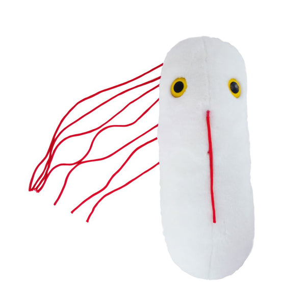


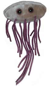


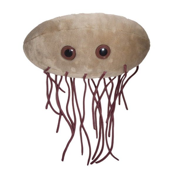


GiantMicrobes är mjukdjur som ser ut som små, små mikrober - bara det att de är förstorade sådär en miljon gånger. De ger bokstavligen ett ansikte åt förkylningen, fotsvampen, hostan, den dåliga andedräkten eller vägglusen. Varje GIGANTmicrobe är mellan ca 38-50 cm. Med följer ett foto på hur den riktiga mikroben ser ut samt en kort information på engelska.
Ursprungligen skapade i USA att användas i undervisningssyfte, har GIANTmicrobes nu blivit storsäljare i museishoppar, apotek, bokhandlar och designbutiker världen över.
GIANTmicrobes är ett roligt verktyg i undervisningen om hälsa och sjukdomar, men även en uppskattad gåva som passar alla åldrar. Definitivt roligare än ett krya-på-dig kort till en sjuk vän.
-------------
en helt vanlig oftast ofalig tarmbakterie. Dessutom forskarnas absoluta favorit!
----------
Escherichia coli
| Escherichia coli | |
|
E. coli
|
|
| Bakterier | |
| Proteobacteria | |
| Gamma Proteobacteria | |
| Enterobacteriales | |
| Enterobacteriaceae | |
| Escherichia | |
| Coli | |
| § Escherichia coli | |
| Theodor Escherich, 1885 | |
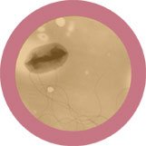
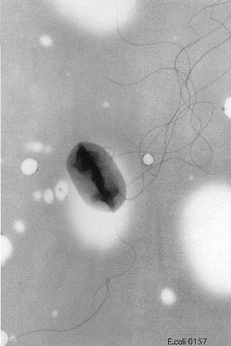
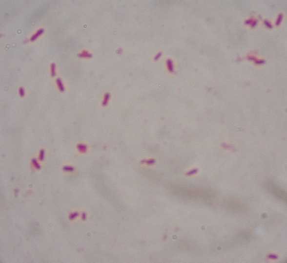
Escherichia coli, förkortas ofta E. coli, är en gramnegativ bakterieart som lever i de nedre delarna av tarmarna hos varmblodiga djur, inklusive fåglar och däggdjur. Det klarlades tidigt att den kan orsaka olika typer av infektioner i bland annat urinvägar, blodbanor, hjärnhinnor och tarmar.
E. coli är nödvändig för normal matsmältning och utgör en stor andel av tarmfloran (intestinala floran). Antalet E. coli-bakterier i en människas avföring varierar mellan 100 miljarder och 10 biljoner per gram feces. Om E. coli påträffas i grundvatten är det troligt att detta förorenats med avföring från människa eller något annat varmblodigt djur.
Maj 2011 skedde ett utbrott av aggressiv E. coli O104:H4 i Tyskland. Senare har även personer i Frankrike och Sverige blivit smittade, med stor utredningsinsats som följd.
Innehåll
[göm]-
1 Morfologi och sjukdomsframkallande egenskaper
- 1.1 Infektioner orsakade av E. coli
- 2 Diagnostik och behandling
- 3 Historia
- 4 Se även
- 5 Källor
Morfologi och sjukdomsframkallande egenskaper[redigera | redigera wikitext]
E. coli ingår i familjen Enterobacteriaceae och studeras ofta som representant för bakterier generellt, särskilt inom molekylärbiologi. E. coli och en mängd andra relativt närbesläktade bakteriarter finns i människans normala tarmflora. Sådana bakterier benämns ofta koliforma bakterier och brukar beskrivas som fakultativt anaeroba rörliga gramnegativa stavar. E. coli växer bra i enkelt sammansatta medier. I näringsbuljong fermenterar de oftast laktos under gasbildning inom 48 timmar vid 35 °C. Hittills har man identifierat 4 olika så kallade ytantigensystem hos E. coli. Dessa benämns O (lipopolysackarid, cirka 190 [1] olika typer finns beskrivna)-, K (kapsel)-, H (flagell)- och F (fimbrie)-antigen. De olika antigenen ger bakterierna en del av deras karakteristiska egenskaper, exempelvis förmåga att orsaka feber (O), motstå kroppens immunförsvar (K), rörlighet (H) och förmåga att fästa på slemhinnor (F). En del av E. coli-bakterierna bildar så kallade enterotoxiner, det vill säga gifter som orsakar diarré. Vissa stammar kan orsaka blödande tarminfektioner och skada njurarna (så kallade EHEC-bakterier, Enterohemorragisk E. coli).
Infektioner orsakade av E. coli[redigera | redigera wikitext]
- Infektioner utanför tarmen: Hjärnhinneinflammation som orsakas av E. coli är mycket ovanlig. Infektionen drabbar i första hand nyfödda. Bakterierna är oftast av K-typ 1. Urinvägsinfektioner är vanligast hos kvinnor och drabbar årligen ungefär 10 % av den kvinnliga befolkningen. Vanligaste orsak är E. coli. K-och F-antigenerna anses ha störst betydelse för bakteriernas förmåga att orsaka urinvägsinfektioner. Vissa bakteriestammar kan ge upphov till infektioner i njurarna (pyelonefrit), ett sjukdomstillstånd som yttrar sig i feber och ryggsmärtor. Infektioner i blodbanorna kan ibland orsakas av E. coli.
- Infektioner i tarmarna: E. coli är den vanligaste orsaken till bakteriell diarré. Man har identifierat 4 olika typer av bakterien som orsak. Dessa benämns ETEC (en typ som är vanlig framförallt i utvecklingsländer och som kan orsaka turistdiarré), EPEC (orsakar diarré framförallt hos småbarn i utvecklingsländer), EIEC (ger upphov till kraftig diarré, förekommer sällan i Sverige), EHEC (har samma slags gift, verotoxin, som Shigella. Infektionerna kan vara allvarliga med bland annat njurpåverkan. De är sällsynta i Sverige).
- Magsjuka kan orsakas ofta av Escherichia coli O157:H7 som finns naturligt på många ställen, som i kons mage. Den dör om den upphettas, men eftersom den sprids så lätt kan den enkelt fästa på t ex en sallad som varit i kontakt med kokött. Sjukdomen i sig orsakas av verocytotoxin som bakterien bildar. En normal infektion varar 5-10 dagar och kan vara smärtsam eller i sällsynta fall, dödlig. Infektionen kan enkelt undvikas med god hygien och tillräckligt upphettad mat.
Diagnostik och behandling[redigera | redigera wikitext]
Enklast diagnostiseras E. coli-infektioner genom att göra en bakteriologisk odling från den infekterade lokalen, till exempel urin eller blod. Eftersom urinvägsinfektioner oftast orsakas av E. coli behöver läkaren ofta inte företa odling av urin, utan kan behandla infektionen med antibiotika ändå.
Infektioner som orsakas av E. coli är behandlingsbara med ett flertal olika antibiotikatyper som pivmecillinam, ampicillin, cefalosporiner, trimetoprim, kinoloner. Mot ampicillin, som är ett slags penicillin, föreligger dock relativt ofta resistens, varför just detta antibiotikum används relativt sällan numera.Klinisk bakteriologi, Arne Forsgren, Göran Kronvall (red.). 1996, Studentlitteratur.
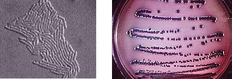
Left: Escherichia coli microcolony on agar surface. Right: E. coli colonies on EMB Agar. On EMB agar E. coli colonies develop a purple color and have a characteristic green sheen. Colonies of Salmonella and Shigella would appear colorless, so the medium can be used to differentiate E. coli from the pathogens.
Rapid Methods for Detecting E. coli
A fluorogenic detection method has been developed based on the cleavage of methylumbelliferyl-D-glucuronide (MUG) to the free methylumbelliferyl moiety, which fluoresces a blue color after irradiation with long-wave ultraviolet radiation. Although strains of E. coli are generally positive in this test, some strains of Salmonella, Shigella, and Yersinia are also capable of splitting MUG; the latter two genera are usually not present in food. A disadvantage is that enterohemorrhagic E. coli (EHEC) strains are generally negative in this test. MUG can be added to various selective media, so there is a great potential in its use for detecting E. coli.
Automated or semi-automated systems are also being used for the detection of E. coli as part of the detection methods for Enterobacteriaceae. Techniques involving impedance measurements have shown promise. Other techniques such as immunoassays and nucleic acid hybridization studies can also be used to enumerate E. coli, and DNA probes directed at a number of genes have also been developed.
Physiology of E. coli
Physiologically, E. coli is versatile and well-adapted to its characteristic habitats. In the laboratory it can grow in media with glucose as the sole organic constituent. Wild-type E. coli has no growth factor requirements, and metabolically it can transform glucose into all of the molecular components that make up the cell. The bacterium can grow in the presence or absence of O2. Under anaerobic conditions it will grow by means of fermentation, producing characteristic "mixed acids and gas" as end products. However, it can also grow by means of anaerobic respiration, since it is able to utilize NO3 or fumarate as final electron acceptors for respiratory electron transport processes. In part, this adapts E. coli to its intestinal (anaerobic) and its extraintestinal (aerobic or anaerobic) habitats.
In the ecological niches that E. coli occupies and its abilities to grow both aerobically and anaerobically are important. E. coli is well adapted to its intestinal environment as it is able to survive on a relatively limited number of low-molecular weight substances, which may only be available transiently and at relatively low concentrations. The generation time for E. coli in the intestine is thought to be about 12 hours. The type of nutrients available there to E. coli consist of mucus, desquamated cells, intestinal enzyme secretions, and incompletely digested food. Given the absorption capacity and efficiency of the intestine, there are probably only small amounts free carbohydrates or other easily absorbable forms of nutrients, and there is competition from hundreds of other types of bacteria. A similar situation probably also applies to sources of nitrogen.
In its natural environment, as well as the laboratory, E. coli can respond to environmental signals such as chemicals, pH, temperature, osmolarity, etc., in a number of very remarkable ways considering it is a single cell organism. For example, it can sense the presence or absence of chemicals and gases in its environment and swim towards or away from them. Or it can stop swimming and grow fimbriae that will specifically attach it to a cell or surface receptor. In response to changes in temperature and osmolarity, it can vary the pore diameter of its outer membrane porins to accommodate larger molecules (nutrients) or to exclude inhibitory substances (e.g. bile salts). With its complex mechanisms for regulation of metabolism the bacterium can survey the chemical content its environment in advance of synthesizing any enzymes necessary to use these compounds. It does not wastefully produce enzymes for degradation of carbon sources unless they are available, and it does not produce enzymes for synthesis of metabolites if they are available as nutrients or growth factors in the environment.
Escherichia coli in the Gastrointestinal Tract
The commensal E. coli strains that inhabit the large intestine of all humans and warm-blooded animals comprise about 1% of the total bacterial biomass. This E. coli flora is in constant flux. One study on the distribution of different E. coli strains colonizing the large intestine of women during a one year period (in a hospital setting) showed that 52.1% yielded one serogroup, 34.9% yielded two, 4.4% yielded three, and 0.6% yielded four. The most likely source of new serotypes of E. coli is acquisition by the oral route. To study oral acquisition, the carriage rate of E. coli carrying antibiotic resistance (R) plasmids was examined among vegetarians, babies, and non vegetarians. It was assumed that non vegetarians might carry more E. coli with R factors due to their presumed high incidence in animals treated with growth-promoting antimicrobial agents. However, omnivores had no higher an incidence of R-factor-containing E. coli than vegetarians, and babies had more resistant E. coli in their feces than non vegetarians. No suitable explanation could be offered for these findings. Besides, investigation of the microbial flora of the uninhabited Krakatoa archipelago has shown the presence of antibiotic-resistant E. coli associated with plants.
Infections Caused by Pathogenic E. coli
E. coli is responsible primarily for three types of infections in humans: urinary tract infections, neonatal meningitis, and intestinal diseases. These conditions depend on a specific array of pathogenic (virulence) determinants possessed by the organism. Pathogenic E. coli are discussed elsewhere in the text in more detail at Pathogenic E. coli: Gastroenteritis, Urinary tract Infections and Neonatal Meningitis.
Urinary Tract Infections
Uropathogenic E. coli cause 90% of the urinary tract infections (UTI) in anatomically-normal, unobstructed urinary tracts. The bacteria colonize from the feces or perineal region and ascend the urinary tract to the bladder. Bladder infections are 14 times more common in females than males by virtue of the shortened urethra. The typical patient with uncomplicated cystitis is a sexually-active female who was first colonized in the intestine with a uropathogenic E. coli strain. The organisms are propelled into the bladder from the periurethral region during sexual intercourse. With the aid of specific fimbriae they are able to colonize the bladder.
The frequency of the distribution of the host cell receptor for the bacterial fimbriae plays a role in susceptibility and explains why certain individuals have repeated UTI caused by E. coli. Uncomplicated E. coli UTI virtually never occurs in individuals lacking the receptors.
Neonatal meningitis
Neonatal meningitis affects 1/2,000-4,000 infants. Eighty percent of E. coli strains involved synthesize K-1 capsular antigens (K-1 is only present 20-40% of the time in intestinal isolates).
E. coli strains invade the blood stream of infants from the nasopharynx or GI tract and are carried to the meninges.
Epidemiological studies have shown that pregnancy is associated with increased rates of colonization by K-1 strains and that these strains become involved in the subsequent cases of meningitis in the newborn. Probably, the infant GI tract is the portal of entry into the bloodstream. Fortunately, although colonization is fairly common, invasion and the catastrophic sequelae are rare.
Neonatal meningitis requires antibiotic therapy that usually includes ampicillin and a third-generation cephalosporin.
Relaterade Produkter