


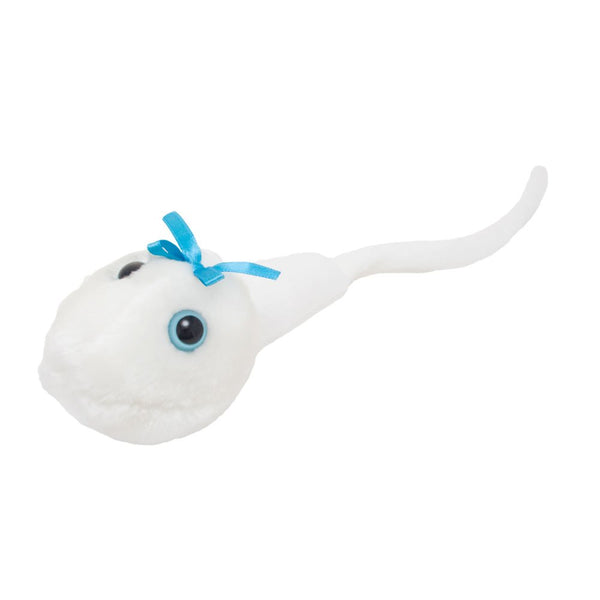


Spermie Cell - Spermatozoon - Sperm - Manlig Könscell
Till någon speciell, någon man älskar eller någon som snart skall ha barn... bara fantasin sätter gränser :-)
Var man har många men här får du blott en :-)
Our little man's man is the stuff of legends. You can bank on it.
Spermie i Mikrosokop:
http://www.youtube.com/watch?v=vvnEsOaKxuw&NR=1
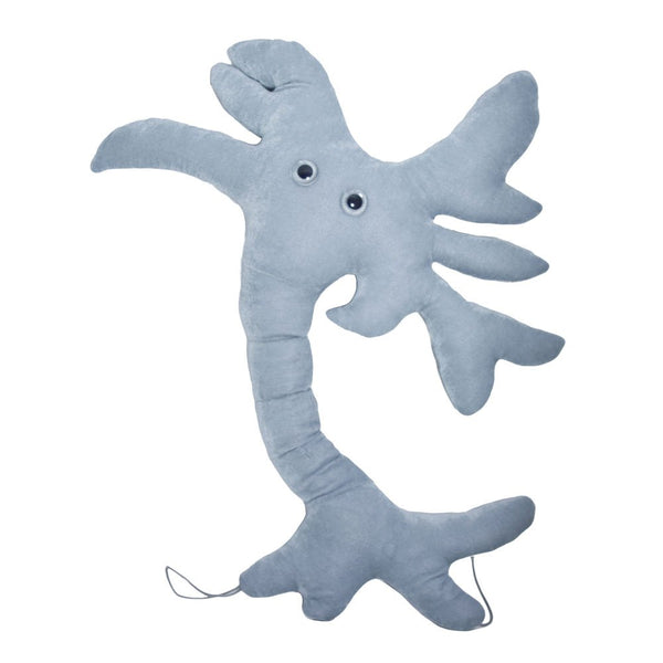
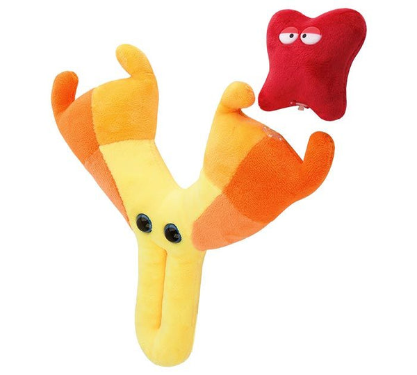
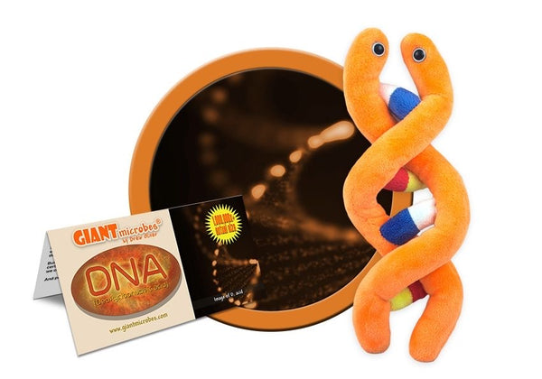
GiantMicrobes är mjukdjur som ser ut som små, små mikrober - bara det att de är förstorade sådär en miljon gånger. De ger bokstavligen ett ansikte åt förkylningen, fotsvampen, hostan, den dåliga andedräkten eller vägglusen. Varje stor GIGANTmicrobe är mellan ca 38-50 cm. De små 10-20cm. Med följer ett foto på hur den riktiga mikroben ser ut samt en kort information på engelska.
Ursprungligen skapade i USA att användas i undervisningssyfte, har GIANTmicrobes nu blivit storsäljare i museishoppar, apotek, bokhandlar och designbutiker världen över.
GIANTmicrobes är ett roligt verktyg i undervisningen om hälsa och sjukdomar, men även en uppskattad gåva som passar alla åldrar. Definitivt roligare än ett krya-på-dig kort till en sjuk vän.
-------------
Spermie
Spermie eller spermatozo är den manliga könscellen, sädescellen. Spermier, vilka bildas i pungen, utsöndras med ett vitt sekret (sperma eller sädesvätska) vid ejakulation, och har till funktion att befrukta äggceller vid sexuell förökning. Produktion av spermier kallas spermatogenes, och sker i testiklarna där runda celler som kallas spermatogonier delar sig och mognar till färdiga spermier.
Innehåll:
Historia[redigera | redigera wikitext]
Ordet spermie kommer från grekiskans sperma, som betyder "säd" och zoon, som betyder "levande". Spermier är små haploida celler, vilket innebär att de inte har en full uppsättning kromosomer. Den förste som observerade och beskrev spermier var Antonie van Leeuwenhoek, vars rön publicerades av Royal Society år 1679.[1]
Beståndsdelar[redigera | redigera wikitext]
En spermie består av ett huvud, mellanstycke och svans. I huvudet finns kromosomerna tätt packade, samt en mindre mängd cellplasma. Längst fram i huvudet finns akrosomen med enzymer som behövs för att kunna tränga igenom äggets skyddande membran, zona pellucida, och befrukta ägget. I mellanstycket finns en stor mängd mitokondrier, som ger den energi spermien behöver för att kunna ta sig fram, genom produktion av ATP. Svansen är en flagell som används för att driva spermien framåt. Denna flagell slår inte från sida till sida utan roterar, på grund av flagellens uppbyggnad med nio microtubilii en ring, och två stycken i mitten[2].
Under befruktningen är det bara kromosomerna som går in i ägget, och därför blir spermiens mitokondrier förstörda av ägget utanför skalet, varför allt mitokondriellt DNA hos barnet är ärvt via modern.
Äggcellen är täckt av ett membran, zona pellucida. När spermien når ägget, frisläpps spermiens enzymer ur akrosomen och bryter ner cellmembranet. Spermiens cellmembran fuserar sedan med äggets, och dess innehåll töms in i ägget.
Människans spermier[redigera | redigera wikitext]
Människans spermier har ett huvud som är sex gånger tre mikrometer stort, och en svans som är 50 mikrometer lång. Den rör sig framåt med ungefär en till tre millimeter per minut.
Spermien innehåller halva det genetiska materialet som behövs för att skapa en ny individ, och ägget den andra hälften. Detta innebär också att endast 50% av mannens anlag förs vidare till avkomman. Människospermien innehåller antingen en X- eller en Y-kromosom, medan ägget alltid innehåller en X-kromosom, och därför är det, hos människan, spermien som avgör vilket kön det blir på barnet.
Spermier bildas kontinuerligt och i astronomiska antal. Det antas att evolutionella faktorer samt tävling mellan olika mäns spermier (spermiekonkurrens) ligger bakom det stora antalet spermier, eftersom detta leder till en tävling mellan spermierna om vilken som kommer att befrukta ägget.[3] För att befruktning ska äga rum krävs dels att spermien kan förflytta sig till platsen för befruktning (äggledaren), dels att spermien klarar miljön som i hög grad står under hormoniell kontroll, dels att spermien förmår tränga in i äggcellen. Till sin hjälp har spermien sädesvätskan som strax efter ejakulationen blir geléliknande för att skydda spermien mot den sura miljön i vagina, varefter sädesvätskan efter ungefär en halvtimme blir mer vätskelik vilken underlättar förflyttningen. Det tar ungefär någon minut innan den första spermien når äggledaren. Det är den spermie med störst rörlighet som hinner dit först, men det är inte nödvändigtvis den snabbaste spermien som befruktar ägget. Spermier kan överleva i upp till fem dagar.[4]
Mannens ålder påverkar sannolikt kvaliteten på spermierna, på samma sätt som kvinnans ålder påverkar äggcellerna. Detta beror troligen på att ökad ålder leder till att fler spermier bär på fler mutationer. Sådana mutationer saknas dock hos äggcellerna.[5] Spermiernas kvalitet försämras, deras antal minskar och rörligheten
Spermatozoon
|
A sperm cell attempts to penetrate an ovum coat to fertilize it.
|
|
|
Diagram of a human spermatozoon
|
|
| spermatozoon | |
| p.1243 | |
| Spermatozoa | |
| Spermatozoa | |
A spermatozoon (pronounced /ˌspɜrmætəˈzoʊən/, alternate spelling spermatozoön; plural spermatozoa; from Ancient Greek: σπέρμα "seed" and Ancient Greek: ζῷον "living being") is a motile sperm cell, or moving form of the haploid cell that is the male gamete. A spermatozoon joins an ovum to form a zygote. (A zygote is a single cell, with a complete set of chromosomes, that normally develops into an embryo.)
Sperm cells contribute approximately half of the nuclear genetic information to the diploid offspring (excluding, in most cases, mitochondrial DNA). In mammals, the sex of the offspring is determined by the sperm cell: a spermatozoon bearing a Y chromosome will lead to a male (XY) offspring, while one bearing an X chromosome will lead to a female (XX) offspring. Sperm cells were first observed by Anton van Leeuwenhoek in 1677.[1]
Contents
[hide]-
1 Mammalian spermatozoan structure, function, and size
- 1.1 Humans
- 1.2 Avoidance of immune system response
-
2 Spermatozoa in other organisms
- 2.1 Animals
- 2.2 Plants, algae and fungi
- 3 Spermatozoa production in mammals
- 4 Spermatozoa activation
- 5 Artificial storage
- 6 History
- 7 References
- 8 External links
Mammalian spermatozoan structure, function, and size[edit]
Humans[edit]
The human sperm cell is the reproductive cell in males and will only survive in warm environments; once it leaves the male body the sperm's survival likelihood is reduced and it may die, thereby decreasing the total sperm quality. Sperm cells come in two types, "female" and "male". Sperm cells that give rise to female (XX) offspring after fertilization differ in that they carry an X-chromosome, while sperm cells that give rise to male (XY) offspring carry a Y-chromosome.
In male humans, sperm cells consists of a flat, disc shaped head 5 µm by 3 µm and a tail 50 µm long.[2] The tail flagellates, which propels the sperm cell (at about 1–3 mm/minute in humans) by whipping in an elliptical cone.[3] Semen has an alkaline nature, and they do not reach full motility (hypermotility) until they reach the vagina where the alkaline pH is neutralized by acidic vaginal fluids. This gradual process takes 20–30 minutes. In this time, fibrinogen from the seminal vesicles forms a clot, securing and protecting the sperm. Just as they become hypermotile, fibrinolysin from the prostate dissolves the clot, allowing the sperm to progress optimally.
The spermatozoon is characterized by a minimum of cytoplasm and the most densely packed DNA known in eukaryotes. Compared to mitotic chromosomes in somatic cells, sperm DNA is at least sixfold more highly condensed.[4]
The specimen contributes with DNA/chromatin, a centriole and perhaps also an oocyte-activating factor (OAF).[5] It may also contribute with paternal messenger RNA (mRNA), also contributing to embryonic development.[5]
-
Electron micrograph of human spermatozoa magnified 3140 times.
-
Sperm cells in the urine sample of a 45 year old male patient who is being followed with the diagnosis of benign prostate hyperplasia.
-
Another image from the same urine sample as with the image on the left.
The human spermatozoon contains over 6000 different proteins.[6]
Avoidance of immune system response[edit]
Glycoprotein molecules on the surface of ejaculated sperm cells are recognized by all human female immune systems, and interpreted as a signal that the cell should not be rejected. The female immune system might otherwise attack sperm in the reproductive tract. The specific glycoproteins coating sperm cells are also utilized by some cancerous and bacterial cells, some parasitic worms, and HIV-infected white blood cells, thereby avoiding an immune response from the host organism.[7]
The blood-testis barrier, maintained by the tight junctions between the Sertoli cells of the seminiferous tubules, prevents communication between the forming spermatozoa in the testis and the blood vessels (and immune cells circulating within them) within the interstitial space. This prevents them from eliciting an immune response. The blood-testis barrier is also important in preventing toxic substances from disrupting spermatogenesis.
Spermatozoa in other organisms[edit]
Animals[edit]
Fertilization relies on spermatozoa for most sexually reproductive animals.
Some species of fruit fly produce the largest known spermatozoon found in nature.[8] Drosophila melanogaster produces sperm that can be up to 1.8 mm, while its relative Drosophila bifurca produce the largest known spermatozoon, measuring over 58 mm in size. In D. melanogaster the entire sperm, tail included, gets incorporated into the oocyte cytoplasm, however, for D. bifurca only a small portion of the tail enters the oocyte.[9]
The wood mouse Apodemus sylvaticus possesses spermatozoa with falciform morphology. What makes these gametocytes even more unique is the presence of an apical hook on the sperm head. This hook is used to attach to the hooks or to the flagella of other spermatozoa. Aggregation is caused by these attachments and mobile trains result. These trains provide improved motility in the female reproductive tract and are a means by which fertilization is promoted.[10]
Sea urchins such as Arbacia punctulata—are ideal organisms to use in sperm research, they spawn large numbers of sperm into the sea, making them well-suited as model organisms for experiments.
Plants, algae and fungi[edit]
The gametophytes of bryophytes, ferns and some gymnosperms produce motile sperm cells, contrary to pollen grains employed in most gymnosperms and all angiosperms. This renders sexual reproduction in the absence of water impossible, since water is a necessary medium for sperm and egg to meet. Algae and lower plant sperm cells are often multi-flagellated (see image) and thus morphologically different from animal spermatozoa.
Some algae and fungi produce non-motile sperm cells, called spermatia. In higher plants and some algae and fungi, fertilization involves the migration of the sperm nucleus through a fertilization tube (e.g. pollen tube in higher plants) to reach the egg cell.
Spermatozoa production in mammals[edit]
Spermatozoa are produced in the seminiferous tubules of the testes in a process called spermatogenesis. Round cells called spermatogonia divide and differentiate eventually to become spermatozoa. During copulation the cloaca or vagina gets inseminated, and then the spermatozoa move through chemotaxis to the ovum inside a Fallopian tube or the uterus.
Spermatozoa activation[edit]
Approaching the egg cell is a rather complex, multistep process of chemotaxis guided by different chemical substances/stimuli on individual levels of phylogeny. One of the most significant, common signaling character of the event is that a prototype of professional chemotaxis receptors, formyl peptide receptor (60.000 receptor/cell) as well as the activator ability of its ligand formyl Met-Leu-Phe have been demonstrated in the surface membrane even in the case of human sperms.[11] Mammalian sperm cells become even more active when they approach an egg cell in a process called sperm activation. Sperm activation has been shown to be caused by calcium ionophores in vitro, progesterone released by nearby cumulus cells and binding to ZP3 of the zona pellucida. The cumulus cells are embedded in a gel-like substance made primarily of hyaluronic acid, and developed in the ovary with the egg and support it as it grows.
The initial change is called "hyperactivation", which causes a change in spermatozoa motility. They swim faster and their tail movements become more forceful and erratic.
A recent discovery links hyperactivation to a sudden influx of calcium ion into the tails. The whip-like tail (flagellum) of the sperm is studded with ion channels formed by proteins called CatSper. These channels are selective, allowing only calcium ion to pass. The opening of CatSper channels is responsible for the influx of calcium. The sudden rise in calcium levels causes the flagellum to form deeper bends, propelling the sperm more forcefully through the viscous environment. Sperm hyperactivity is necessary for breaking through two physical barriers that protect the egg from fertilization.
The second process in sperm activation is the acrosome reaction. This involves releasing the contents of the acrosome, which disperse, and the exposure of enzymes attached to the inner acrosomal membrane of the sperm. This occurs after the sperm first meets the egg. This lock-and-key type mechanism is species-specific and prevents the sperm and egg of different species from fusing. There is some evidence that this binding is what triggers the acrosome to release the enzymes that allow the sperm to fuse with the egg.
ZP3, one of the proteins that make up the zona pellucida, then binds to a partner molecule on the sperm. Enzymes on the inner acrosomal membrane digests the zona pellucida. After the sperm penetrates the zona pellucida, part of the sperm's cell membrane then fuses with the egg cell's membrane, and the contents of the head diffuse into the egg.
Upon penetration, the oocyte is said to have become activated. It undergoes its secondary meiotic division, and the two haploid nuclei (paternal and maternal) fuse to form a zygote. In order to prevent polyspermy and minimise the possibility of producing a triploid zygote, several changes to the egg's zona pellucida renders them impenetrable shortly after the first sperm enters the egg.
Artificial storage[edit]
Spermatozoa can be stored in diluents such has the Illini Variable Temperature (IVT) diluent, which have been reported to be able to preserve high fertility of spermatozoa for over seven days.[12] The IVT diluent is composed of several salts, sugars and antibacterial agents and gassed with CO2.[12]
Semen cryopreservation can be used for far longer storage durations. For human spermatozoa, the longest reported successful storage with this method is 21 years.[13]
History[edit]
- In 1677 microbiologist Antonie van Leeuwenhoek discovers spermatozoa.
- In 1841 the Swiss anatomist Albert von Kölliker wrote about spermatozoon in his work Untersuchungen uber die Bedeutung der Samenfäden.
References[edit]
- Jump up ^ "Timeline: Assisted reproduction and birth control". CBC News. Retrieved 2006-04-06.
- Jump up ^ Smith, D.J. (2009). "Human sperm accumulation near surfaces: a simulation study". Journal of Fluid Mechanics 621: 295. doi:10.1017/S0022112008004953. Retrieved 20 May 2012.
- Jump up ^ Sumio Ishijima, Shigeru Oshio, Hideo Mohri, "Flagellar movement of human spermatozoa", Gamete research, 1986, vol. 13, no3, pp. 185–197 (27 ref.) [1]
- Jump up ^ Ward WS, Coffey DS (1991). "DNA packaging and organization in mammalian spermatozoa: comparison with somatic cells". Biol. Reprod. 44 (4): 569–74. doi:10.1095/biolreprod44.4.569. PMID 2043729.
- ^ Jump up to: a b Developmental sperm contributions: fertilization and beyond Gerardo Barroso, M.D., M.Sc.a, Carlos Valdespin, M.D.a, Eva Vega, M.Sc.a, Ruben Kershenovich, M.D.a, Rosaura Avila, B.Sc.a, Conrado Avendaño, M.D.b, Sergio Oehninger, M.D., Ph.D.b. FertStert, Volume 92, Issue 3, Pages 835-848 (September 2009)
- Jump up ^ Amaral, A.; Castillo, J.; Ramalho-Santos, J.; Oliva, R. (2013). "The combined human sperm proteome: Cellular pathways and implications for basic and clinical science". Human Reproduction Update 20: 40. doi:10.1093/humupd/dmt046. edit
- Jump up ^ "Sperm clue to 'disease immunity'". BBC News. 2007-12-17.
- Jump up ^ Than, Ker. "Longest Known Sperm Create Paradox of Nature". www.livescience.com. Retrieved 20 May 2012.
- Jump up ^ Pitnick, S., Spicer, G. S., & Markow, T. A. (1995). How long is a giant sperm Nature, 375(6527), 109. doi:10.1038/375109a0
- Jump up ^ Moore, Harry et al., Exceptional sperm cooperation in Wood Mouse. Nature 418, 174–177 (2002)
- Jump up ^ Gnessi L, Fabbri A, Silvestroni L, Moretti C, Fraioli F, Pert CB, Isidori A. (1986). "Evidence for the presence of specific receptors for N-formyl chemotactic peptides on human spermatozoa.". J Clin Endocrinol Metab 63 (4): 841–6. doi:10.1210/jcem-63-4-841. PMID 3018025
- ^ Jump up to: a b Watson, P. F. (1993). "The potential impact of sperm encapsulation technology on the importance of timing of artificial insemination: A perspective in the light of published work". Reproduction, Fertility and Development 5 (6): 691–9. doi:10.1071/RD9930691. PMID 9627729. edit
- Jump up ^ Planer NEWS and Press Releases > Child born after 21 year semen storage using Planer controlled rate freezer 14/10/2004
External links[edit]
| Wikimedia Commons has media related to Spermatozoon diagrams. |
- Slower conception 'leads to boys'
- Human Sperm Under a Microscope