


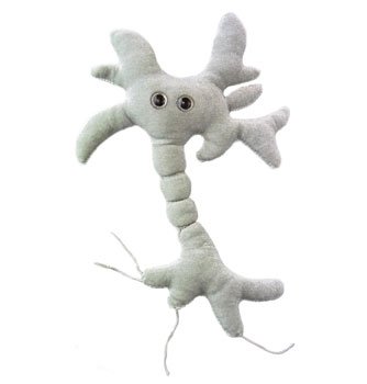


GiantMicrobes är mjukdjur som ser ut som små, små mikrober - bara det att de är förstorade sådär en miljon gånger. De ger bokstavligen ett ansikte åt förkylningen, fotsvampen, hostan, den dåliga andedräkten eller vägglusen. Varje stor GIGANTmicrobe är mellan ca 38-50 cm. De små ca.10-20cm. Med följer ett foto på hur den riktiga mikroben ser ut samt en kort information på engelska.
Ursprungligen skapade i USA att användas i undervisningssyfte, har GIANTmicrobes nu blivit storsäljare i museishoppar, apotek, bokhandlar och designbutiker världen över.
GIANTmicrobes är ett roligt verktyg i undervisningen om hälsa och sjukdomar, men även en uppskattad gåva som passar alla åldrar. Definitivt roligare än ett krya-på-dig kort till en sjuk vän.
-------------
---------
Är du smart eller???
Kanske en bra present till Studenten eller disputationen!
................
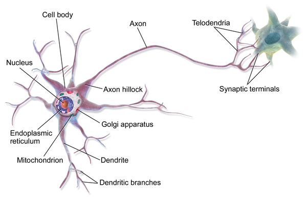
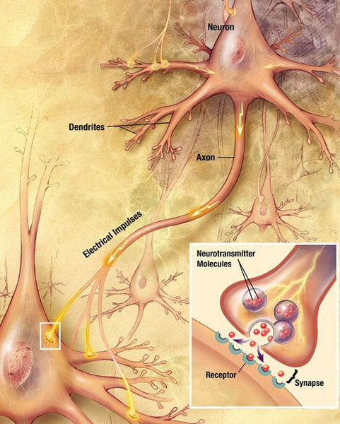
Nervcell (Hjärncell)
Nervcell
1. Korniga endoplasmatiska nätverket (Nisslkropp) 2. Polysomer 3. Ribosomer 4. Golgiapparaten 5. Cellkärna 6. Nukleol 7. Cellmembran 8. Mikrotubuli 9. Mitokondrie 10. Glatta endoplasmatiska nätverket 11. Axonkägla 12. Cellkärna (Schwanncell) 13. Synaps (axosomatisk) 14. Synapser (axodendritiska) 15. Dendriter 16. Axon 17. Neurotransmittor 18. Receptor 19. Synaps 20. Cellskelett 21. Myelinskida (Schwanncell) 22. Ranviernod 23. Axonterminal 24. Synapsvesikel 25. Synaps (axoaxonisk) 26. Synapsspalt
Overview[edit]
Structure of a typical neuronNeuron (peripheral nervous system)|
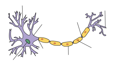
Dendrite
Soma
Axon
Nucleus
Node of Ranvier
Axon terminal
Schwann cell
Myelin sheath
|
A neuron is a specialized type of cell found in the bodies of all eumetozoans. Only sponges and a few other simpler animals lack neurons. The features that define a neuron are electrical excitability and the presence of synapses, which are complex membrane junctions that transmit signals to other cells. The body's neurons, plus the glial cells that give them structural and metabolic support, together constitute the nervous system. In vertebrates, the majority of neurons belong to the central nervous system, but some reside in peripheral ganglia, and many sensory neurons are situated in sensory organs such as the retina and cochlea.
Although neurons are very diverse and there are exceptions to nearly every rule, it is convenient to begin with a schematic description of the structure and function of a "typical" neuron. A typical neuron is divided into three parts: the soma or cell body, dendrites, and axon. The soma is usually compact; the axon and dendrites are filaments that extrude from it. Dendrites typically branch profusely, getting thinner with each branching, and extending their farthest branches a few hundred micrometers from the soma. The axon leaves the soma at a swelling called the axon hillock, and can extend for great distances, giving rise to hundreds of branches. Unlike dendrites, an axon usually maintains the same diameter as it extends. The soma may give rise to numerous dendrites, but never to more than one axon. Synaptic signals from other neurons are received by the soma and dendrites; signals to other neurons are transmitted by the axon. A typical synapse, then, is a contact between the axon of one neuron and a dendrite or soma of another. Synaptic signals may be excitatory or inhibitory. If the net excitation received by a neuron over a short period of time is large enough, the neuron generates a brief pulse called an action potential, which originates at the soma and propagates rapidly along the axon, activating synapses onto other neurons as it goes. This is called saltatory conduction.
Many neurons fit the foregoing schema in every respect, but there are also exceptions to most parts of it. There are no neurons that lack a soma, but there are neurons that lack dendrites, and others that lack an axon. Furthermore, in addition to the typical axodendritic and axosomatic synapses, there are axoaxonic (axon-to-axon) and dendrodendritic (dendrite-to-dendrite) synapses.
The key to neural function is the synaptic signaling process, which is partly electrical and partly chemical. The electrical aspect depends on properties of the neuron's membrane. Like all animal cells, the cell body of every neuron is enclosed by a plasma membrane, a bilayer of lipid molecules with many types of protein structures embedded in it. A lipid bilayer is a powerful electrical insulator, but in neurons, many of the protein structures embedded in the membrane are electrically active. These include ion channels that permit electrically charged ions to flow across the membrane, and ion pumps that actively transport ions from one side of the membrane to the other. Most ion channels are permeable only to specific types of ions. Some ion channels are voltage gated, meaning that they can be switched between open and closed states by altering the voltage difference across the membrane. Others are chemically gated, meaning that they can be switched between open and closed states by interactions with chemicals that diffuse through the extracellular fluid. The interactions between ion channels and ion pumps produce a voltage difference across the membrane, typically a bit less than 1/10 of a volt at baseline. This voltage has two functions: first, it provides a power source for an assortment of voltage-dependent protein machinery that is embedded in the membrane; second, it provides a basis for electrical signal transmission between different parts of the membrane.
Neurons communicate by chemical and electrical synapses in a process known as neurotransmission also called synaptic transmission. The fundamental process that triggers the release of neurotransmitters is the action potential, a propagating electrical signal that is generated by exploiting the electrically excitable membrane of the neuron. This is also known as a wave of depolarization.
Anatomy and histology[edit]
Neurons are highly specialized for the processing and transmission of cellular signals. Given their diversity of functions performed in different parts of the nervous system, there is, as expected, a wide variety in their shape, size, and electrochemical properties. For instance, the soma of a neuron can vary from 4 to 100 micrometers in diameter.[3]
- The soma is the body of the neuron. As it contains the nucleus, most protein synthesis occurs here. The nucleus can range from 3 to 18 micrometers in diameter.[4]
- The dendrites of a neuron are cellular extensions with many branches. This overall shape and structure is referred to metaphorically as a dendritic tree. This is where the majority of input to the neuron occurs via the dendritic spine.
- The axon is a finer, cable-like projection that can extend tens, hundreds, or even tens of thousands of times the diameter of the soma in length. The axon carries nerve signals away from the soma (and also carries some types of information back to it). Many neurons have only one axon, but this axon may—and usually will—undergo extensive branching, enabling communication with many target cells. The part of the axon where it emerges from the soma is called the axon hillock. Besides being an anatomical structure, the axon hillock is also the part of the neuron that has the greatest density of voltage-dependent sodium channels. This makes it the most easily excited part of the neuron and the spike initiation zone for the axon: in electrophysiological terms it has the most negative action potential threshold. While the axon and axon hillock are generally involved in information outflow, this region can also receive input from other neurons.
- The axon terminal contains synapses, specialized structures where neurotransmitter chemicals are released to communicate with target neurons.
Although the canonical view of the neuron attributes dedicated functions to its various anatomical components, dendrites and axons often act in ways contrary to their so-called main function.
Axons and dendrites in the central nervous system are typically only about one micrometer thick, while some in the peripheral nervous system are much thicker. The soma is usually about 10–25 micrometers in diameter and often is not much larger than the cell nucleus it contains. The longest axon of a human motoneuron can be over a meter long, reaching from the base of the spine to the toes. Sensory neurons have axons that run from the toes to the dorsal columns, over 1.5 meters in adults. Giraffes have single axons several meters in length running along the entire length of their necks. Much of what is known about axonal function comes from studying the squid giant axon, an ideal experimental preparation because of its relatively immense size (0.5–1 millimeters thick, several centimeters long).
Fully differentiated neurons are permanently postmitotic;[5] however, recent research shows that additional neurons throughout the brain can originate from neural stem cells found throughout the brain but in particularly high concentrations in the subventricular zone and subgranular zone through the process of neurogenesis.[6]
Relaterade Produkter