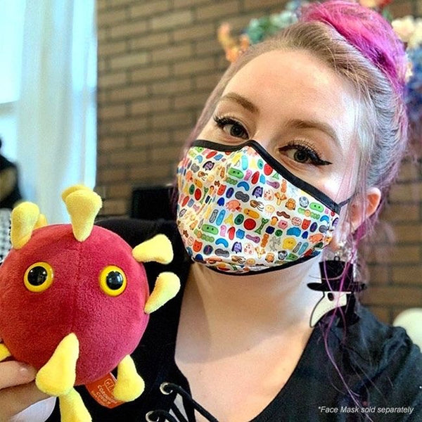


GiantMicrobes är mjukdjur som ser ut som små, små mikrober - bara det att de är förstorade sådär en miljon gånger. De ger bokstavligen ett ansikte åt förkylningen, fotsvampen, hostan, den dåliga andedräkten eller vägglusen. Varje GIGANTmicrobe är mellan ca 38-50 cm. Med följer ett foto på hur den riktiga mikroben ser ut samt en kort information på engelska.
Ursprungligen skapade i USA att användas i undervisningssyfte, har GIANTmicrobes nu blivit storsäljare i museishoppar, apotek, bokhandlar och designbutiker världen över.
GIANTmicrobes är ett roligt verktyg i undervisningen om hälsa och sjukdomar, men även en uppskattad gåva som passar alla åldrar. Definitivt roligare än ett krya-på-dig kort till en sjuk vän.
-------------
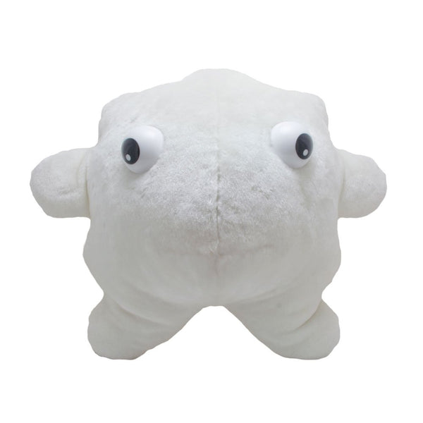
Vita blodkroppar eller leukocyter är celler i blodet som ingår i kroppens immunförsvar för att skydda kroppen mot infektionssjukdomar. Vita blodkroppar produceras i benmärgen och återfinns i lymfknutarna, mjälten och i lungornas alveoler, samt ute i vävnaderna vid inflammatoriska tillstånd. Halten av olika vita blodkroppar kan mätas i ett blodprov för att se om en inflammatorisk process pågår eller om immunförsvaret är nedsatt.
Bara några procent av de vita blodkropparna finns i våra blodkärl, de flesta utanför blodkärlen i kroppens olika vävnader. Deras uppgift i immunförsvaret är att skydda kroppen och ta hand om bakterier, virus och andra ämnen som är främmande för vår kropp.
Referensvärde[redigera | redigera wikitext]
Vid provtagning för medicinskt bruk tar man vanligtvis B-LPK med ett referensvärde för vuxna över 16 år på 4,0-10,0 x 10⁹ vita blodkroppar/liter. [1]
Typer[redigera | redigera wikitext]
Det finns flera olika typer av vita blodkroppar, och en vanlig indelning är i granulocyter och agranulocyter.
| Typ | Bild | Diagram | % av de vita blodkropparna | Beskrivning |
| Neutrofiler | 54-62% | Neutrofiler hanterar försvaret mot bakterieinfektioner och andra mycket små inflammationsprocesser, och är vanligtvis de första försvararna mot bakterieangrepp. Det var som uppstår vid sårläkning består till stor del av döda neutrofiler. | ||
| Eosinofil | 1-6% | Eosinofiler produceras vid infektioner orsakade av endoparasiter och en ökning av antalet eosinofiler kan indikera en sådan infektion. | ||
| Mastcell | [[]] | [[]] | Mastceller bekämpar parasiter, speciellt maskar som infekterat kroppen [[]] Mastceller finns i bindväv och är långsmal med rund kärna och kontrasterande basofil granulae i cytoplasman. Cellmembranet är veckat och det har mikrovilli. | |
| Basofil | <1% | Basofiler ansvarar huvudsakligen för att hantera allergier och rekryteras till vävnad där antigen påträffas. Genom frisättning av histamin stimuleras inflammationen i den angripna vävnaden. | ||
| Lymfocyt | 25-33% |
Lymfocyter är vanliga i det lymfatiska systemet. Blodet innehåller tre typer av lymfocyter:
|
||
| Monocyt | 2-8% | Monocyterna har liksom neutrofilerna "storätar-egenskaper" (fagocytos), men är mer långlivade, då de har ytterligare en roll: de uppvisar bitar av patogen för de patrullerande T-cellerna så att patogenen kan kännas igen och bli oskadliggjorda igen, eller se till att en antikroppsåtgärd aktiveras. | ||
| Makrofag | (se ovan) | Monocyter kallas makrofager efter att de vandrat över från blodomloppet och in i vävnaden. | ||
| Dendritisk cell | Dendritiska celler är professionella antigenpresenterande celler. Dessa fagocyterar främmande ämnen, inkräktande mikroorganismer (etc.) och presenterar dessa för T-hjälparceller. |
White blood cell
|
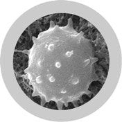
A scanning electron microscope image of normal circulating human blood. In addition to the irregularly shaped leukocytes, both red blood cells and many small disc-shaped platelets are visible.
| |
| leucocytus | |
| TH H2.00.04.1.02001 | |
White blood cells, or leukocytes (also spelled "leucocytes"), are the cells of the immune system that are involved in defending the body against both infectious disease and foreign materials. Five[1] different and diverse types of leukocytes exist, and several types (including monocytes and neutrophils) are phagocytic. All leukocytes are produced and derived from a multipotent cell in the bone marrow known as a hematopoietic stem cell. They live for about three to four days in the average human body. Leukocytes are found throughout the body, including the blood and lymphatic system.[2]
The number of leukocytes in the blood is often an indicator of disease. In the U.S. there are normally approximately 7000 white blood cells per microliter of blood.[3] They make up approximately 1% of the total blood volume in a healthy adult.[4] An increase in the number of leukocytes over the upper limits is called leukocytosis, and a decrease below the lower limit is called leukopenia. Physical properties of leukocytes (such as volume, conductivity, and granularity) may change. These changes can be due to activation, the presence of immature cells, or the presence of malignant leukocytes in leukemia.
Contents
[hide]- 1 Etymology
-
2 Types
- 2.1 Overview
- 2.2 Neutrophil
- 2.3 Eosinophil
- 2.4 Basophil
- 2.5 Lymphocyte
- 2.6 Monocyte
- 3 Fixed leukocytes
-
4 Disorders
-
4.1 Leukopenias
- 4.1.1 Neutropenia
- 4.1.2 Lymphocytopenia
-
4.2 Proliferative disorders
- 4.2.1 Neutrophilia
- 4.2.2 Eosinophilia
-
4.1 Leukopenias
- 5 See also
- 6 References
- 7 External links
Etymology[edit]
The name "white blood cell" derives from the physical appearance of a blood sample after centrifugation. White cells are found in the buffy coat, a thin, typically white layer of nucleated cells between the sedimented red blood cells and the blood plasma. The scientific term leukocyte directly reflects its description. It is derived from the Greek word leuko- meaning "white" and kytos meaning "hollow vessel", with -cyte translated as "cell" in modern usage. Buffy coat may sometimes be green if there are large amounts of neutrophils in the sample, due to the heme-containing enzyme myeloperoxidase that they produce.
Types[edit]
There are several different types of white blood cells. They all have many things in common, but all are distinct in form and function. A major distinguishing feature of some leukocytes is the presence of granules; white blood cells are often characterized as granulocytes or agranulocytes:
- Granulocytes (polymorphonuclear leukocytes): leukocytes characterized by the presence of differently staining granules in their cytoplasm when viewed under light microscopy. These granules (usually lysozymes) are membrane-bound enzymes that act primarily in the digestion of endocytosed particles. There are three types of granulocytes: neutrophils, basophils, and eosinophils, which are named according to their staining properties.
- Agranulocytes (mononuclear leukocytes): leukocytes characterized by the apparent absence of granules in their cytoplasm. Although the name implies a lack of granules these cells do contain non-specific azurophilic granules, which are lysosomes.[5] The cells include lymphocytes, monocytes, and macrophages.[6]
Overview[edit]
TypeMicroscopic AppearanceDiagramApprox. %in adults
See also:
Blood valuesDiameter (μm)[7]Main targets[4]Nucleus[4]Granules[4]Lifetime[7]
| Neutrophil | 62% | 10–12 |
|
Multilobed | Fine, faintly pink (H&E stain) | 6 hours–few days (days in spleen and other tissue) |
||
| Eosinophil | 2.3% | 10–12 |
|
Bi-lobed | Full of pink-orange (H&E stain) | 8–12 days (circulate for 4–5 hours) | ||
| Basophil | 0.4% | 12–15 |
|
Bi-lobed or tri-lobed | Large blue | A few hours to a few days | ||
| Lymphocyte | 30% | Small lymphocytes 7–8 Large lymphocytes 12–15 |
|
Deeply staining, eccentric | NK-cells and cytotoxic (CD8+) T-cells | Years for memory cells, weeks for all else. | ||
| Monocyte | 5.3% | 12–20[8] | Monocytes migrate from the bloodstream to other tissues and differentiate into tissue resident macrophages, Kupffer cells in the liver. | Kidney shaped | None | Hours to days |
Neutrophil[edit]
Neutrophils defend against bacterial or fungal infection. They are usually first responders to microbial infection; their activity and death in large numbers forms pus. They are commonly referred to as polymorphonuclear (PMN) leukocytes, although, in the technical sense, PMN refers to all granulocytes. They have a multi-lobed nucleus that may appear like multiple nuclei, hence the name polymorphonuclear leukocyte. The cytoplasm may look transparent because of fine granules that are pale lilac. Neutrophils are active in phagocytosing bacteria and are present in large amount in the pus of wounds. These cells are not able to renew their lysosomes (used in digesting microbes) and die after having phagocytosed a few pathogens.[9] Neutrophils are the most common cell type seen in the early stages of acute inflammation. They make up 60-70% of total leukocyte count in human blood.[4] The life span of a circulating human neutrophil is about 5.4 days.[10]
Eosinophil[edit]
Eosinophils primarily deal with parasitic infections. Eosinophils are also the predominant inflammatory cells in allergic reactions. The most important causes of eosinophilia include allergies such as asthma, hay fever, and hives; and also parasitic infections. In general, their nucleus is bi-lobed. The cytoplasm is full of granules that assume a characteristic pink-orange color with eosin stain.
Basophil[edit]
Basophils are chiefly responsible for allergic and antigen response by releasing the chemical histamine causing vasodilation. The nucleus is bi- or tri-lobed, but it is hard to see because of the number of coarse granules that hide it. They are characterized by their large blue granules.
Lymphocyte[edit]
Lymphocytes are much more common in the lymphatic system than in blood. Lymphocytes are distinguished by having a deeply staining nucleus that may be eccentric in location, and a relatively small amount of cytoplasm. Lymphocytes include:
- B cells make antibodies that can bind to pathogens, block pathogen invasion, activate the complement system, and enhance pathogen destruction.
-
T cells:
- CD4+ helper T cells: T cells displaying co-receptor CD4 are known as CD4+ T cells. These cells have T-cell receptors and CD4 molecules that, in combination, bind antigenic peptides presented on major histocompatibility complex (MHC) class II molecules on antigen-presenting cells. Helper T cells make cytokines and perform other functions that help coordinate the immune response. In HIV infection, these T cells are the main index to identify the individual's immune system integrity.
- CD8+ cytotoxic T cells: T cells displaying co-receptor CD8 are known as CD8+ T cells. These cells bind antigens presented on MHC I complex of virus-infected or tumour cells and kill them. Nearly all nucleated cells display MHC I.
- γδ T cells possess an alternative T cell receptor (different from the αβ TCR found on conventional CD4+ and CD8+ T cells). Found in tissue more commonly than in blood, γδ T cells share characteristics of helper T cells, cytotoxic T cells, and natural killer cells.
- Natural killer cells are able to kill cells of the body that do not display MHC class I molecules, or display stress markers such as MHC class I polypeptide-related sequence A (MIC-A). Decreased expression of MHC class I and up-regulation of MIC-A can happen when cells are infected by a virus or become cancerous.
Monocyte[edit]
Monocytes share the "vacuum cleaner" (phagocytosis) function of neutrophils, but are much longer lived as they have an extra role: they present pieces of pathogens to T cells so that the pathogens may be recognized again and killed. This causes an antibody response to be mounted. Monocytes eventually leave the bloodstream and become tissue macrophages, which remove dead cell debris as well as attacking microorganisms. Neither dead cell debris nor attacking microorganisms can be dealt with effectively by the neutrophils. Unlike neutrophils, monocytes are able to replace their lysosomal contents and are thought to have a much longer active life. They have the kidney shaped nucleus and are typically agranulated. They also possess abundant cytoplasm.
Once monocytes move from the bloodstream out into the body tissues, they undergo changes (differentiate) allowing phagocytosis and are then known as macrophages.
Fixed leukocytes[edit]
Some leukocytes migrate into the tissues of the body to take up a permanent residence at that location rather than remaining in the blood. Often these cells have specific names depending upon which tissue they settle in, such as fixed macrophages in the liver, which become known as Kupffer cells. These cells still serve a role in the immune system.
- Histiocytes
- Dendritic cells (Although these will often migrate to local lymph nodes upon ingesting antigens)
- Mast cells
- Microglia
Relaterade Produkter